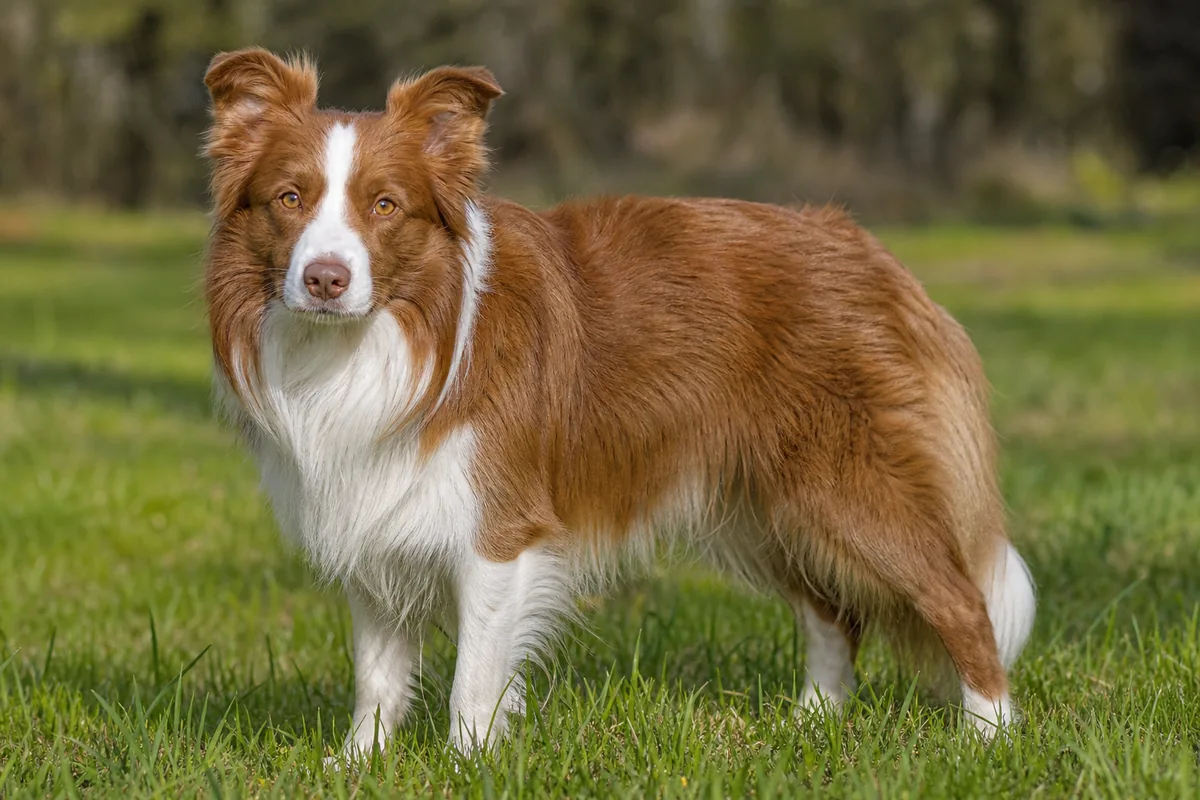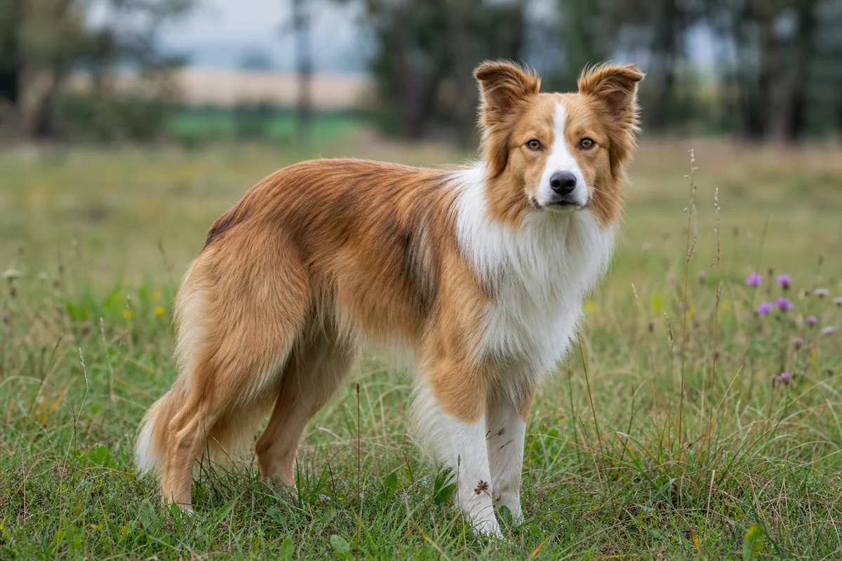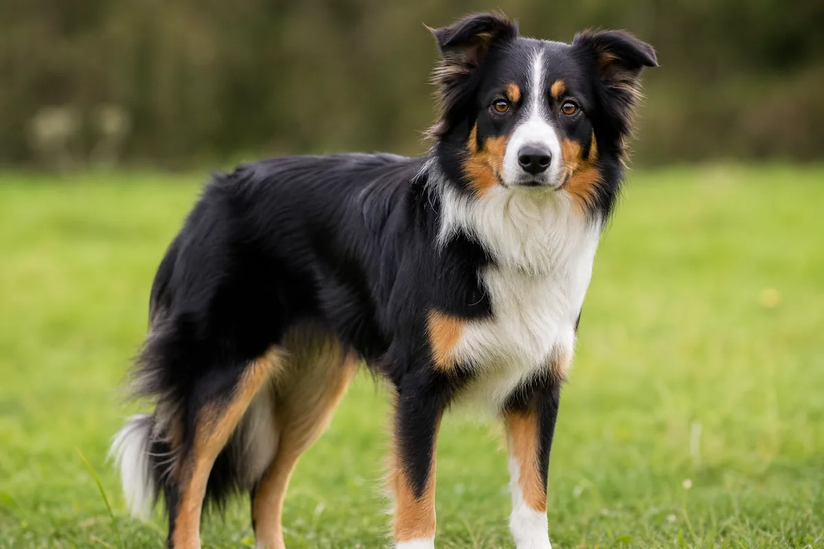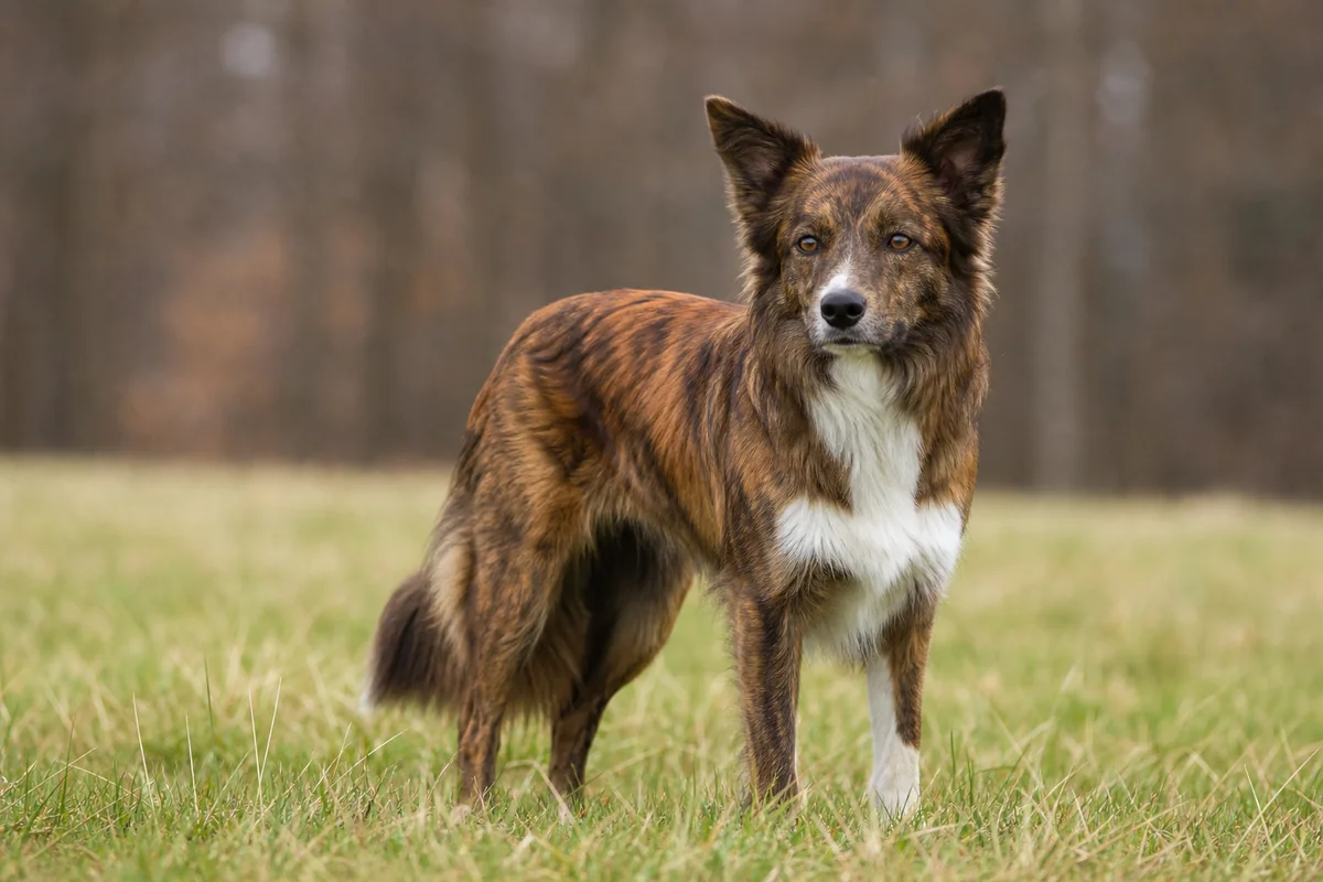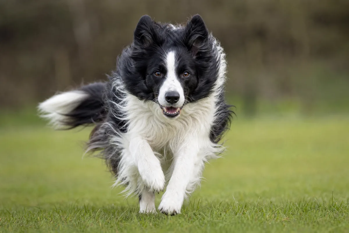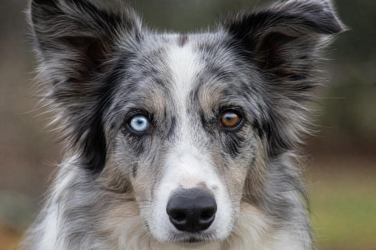
Colour is a description, not a diagnosis. The iris, nose leather, lips and eyelid margins all contain pigment, yet they do not follow one universal “matching” rule. Record what is visible, the dog's age and whether the feature is stable; use genetic reports and veterinary examinations for the different questions they can actually answer.
Iris colour and heterochromia: useful terms
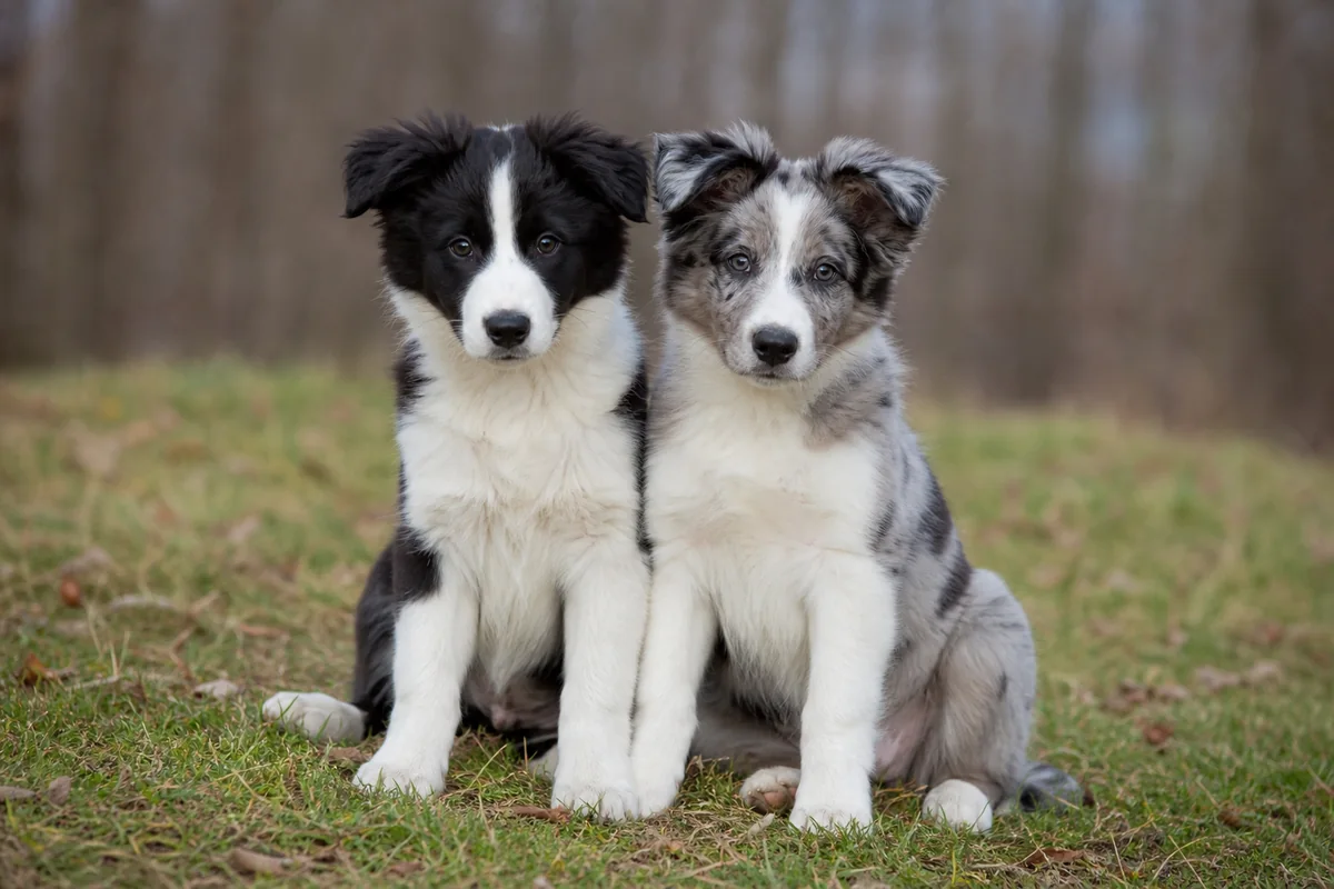
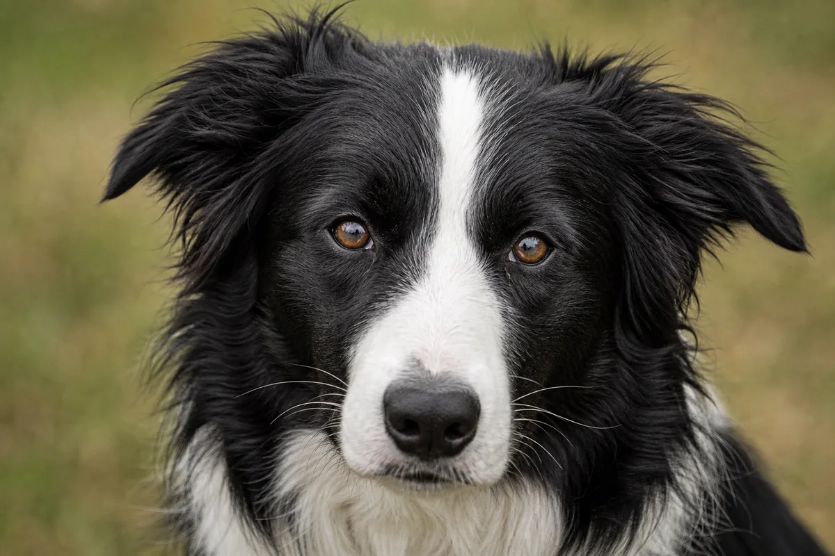
Perceived iris colour reflects pigment amount and distribution within the iris, plus how light interacts with its tissues. Border Collie eyes may look dark brown, medium brown, amber, greenish, grey or blue, with continuous shades between those labels. A pale iris is not transparent, and colour alone says nothing certain about visual function.
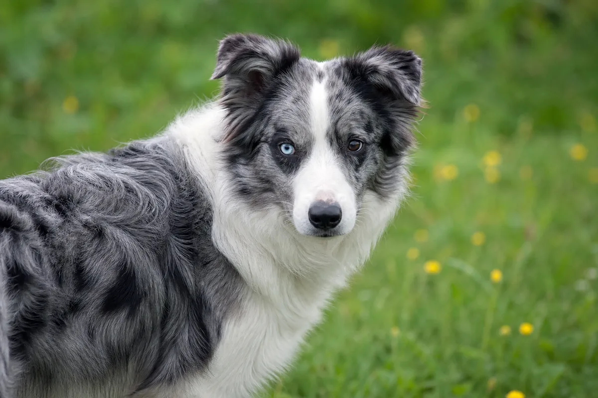
Complete heterochromia means the two irises appear different colours. Sectoral heterochromia means one iris contains differently coloured sectors; people also say “part eye” or “marbled eye.” These are descriptions of pigment distribution. A stable pattern present from puppyhood can be a normal feature. It does not by itself diagnose merle, deafness, blindness or an ocular disorder.
Age, light and photographs change the impression
Puppy pigment can continue to mature, so a young dog's apparent shade should not be treated as a fixed adult result. In any dog, pupil size changes how much iris is visible. Sun, shade, flash, reflections, camera exposure, white balance and image processing can make the same eye look different. A wet nose also reflects more light than a dry surface.
For a descriptive record, use indirect daylight without flash, photograph both eyes and both sides of the nose, and include the date and age. Repeat from the same distance if following a feature. Photos can document change for a veterinarian, but cannot reveal pressure inside the eye, the full lens or retina, microscopic inflammation, tear production, hearing, or a DNA sequence.
How coat-colour pathways relate—and where they stop
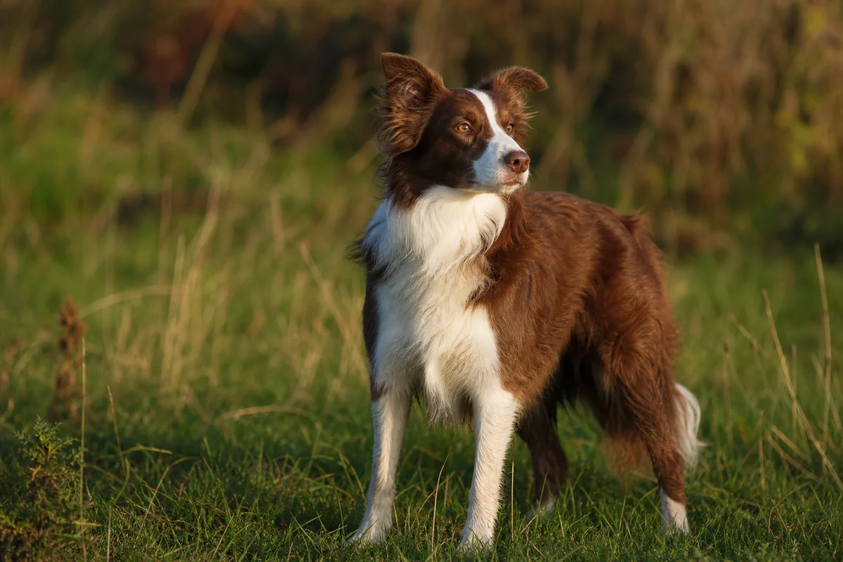
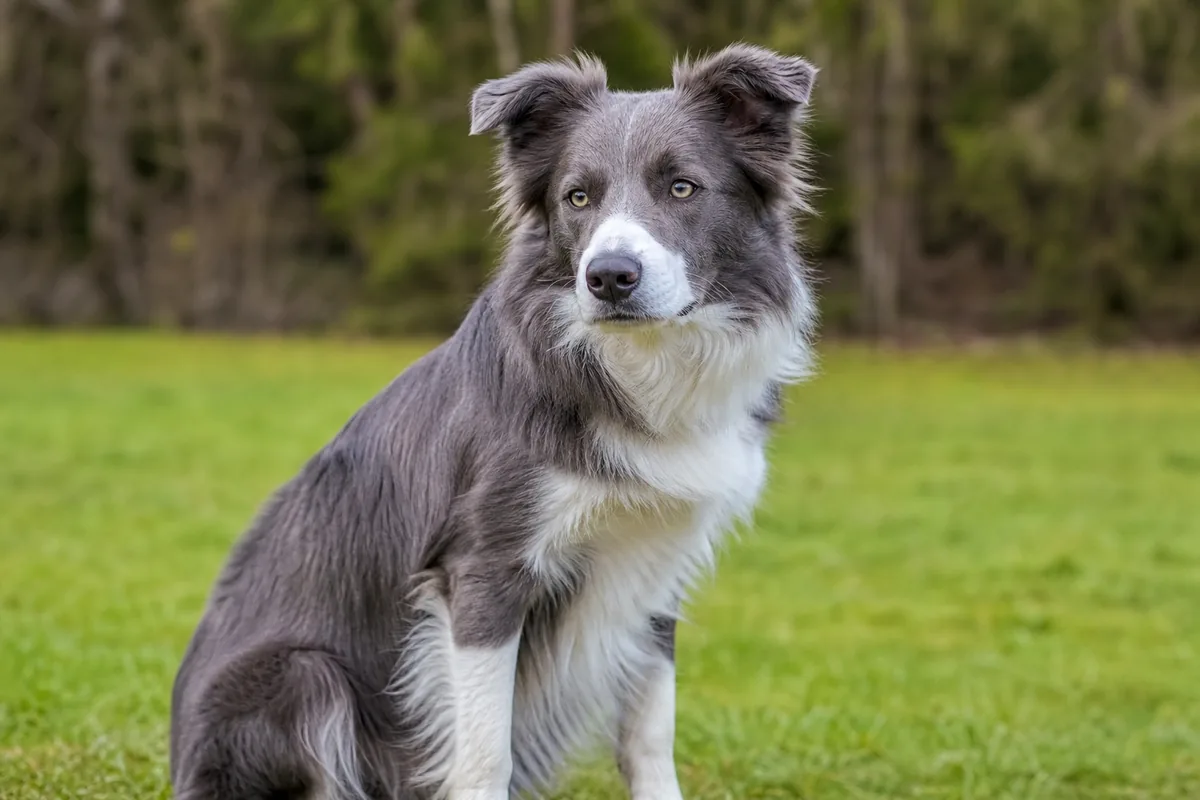
The UC Davis brown test covers named variants in TYRP1 that change black eumelanin to brown. When that pathway is expressed, the nose, lips and eyelid margins may also be brown rather than black. The UC Davis dilution resource describes known MLPH variants that lighten eumelanin, including external pigment. These relationships can make nose colour compatible with a coat hypothesis, but the nose is not a substitute for a genotype result, and neither locus alone predicts a precise iris shade.
Merle is a separate PMEL-related pattern affecting eumelanin. It can be accompanied by blue or partly blue irises and patchy nose pigment, but expression varies. The underlying insertion can also be visually subtle or masked. A primary study of PMEL insertion length demonstrates why visible pattern and molecular result are not interchangeable.
Nor does every blue eye imply PMEL. A large canine genome-wide association study identified a different region near ALX4, primarily associated with blue eyes and heterochromia in Siberian Huskies. That result is not a Border Collie diagnostic test; it illustrates that similar eye colours may have different genetic backgrounds. White spotting, developmental variation and mechanisms not captured by a selected panel add further uncertainty.
Nose leather and eyelid margins
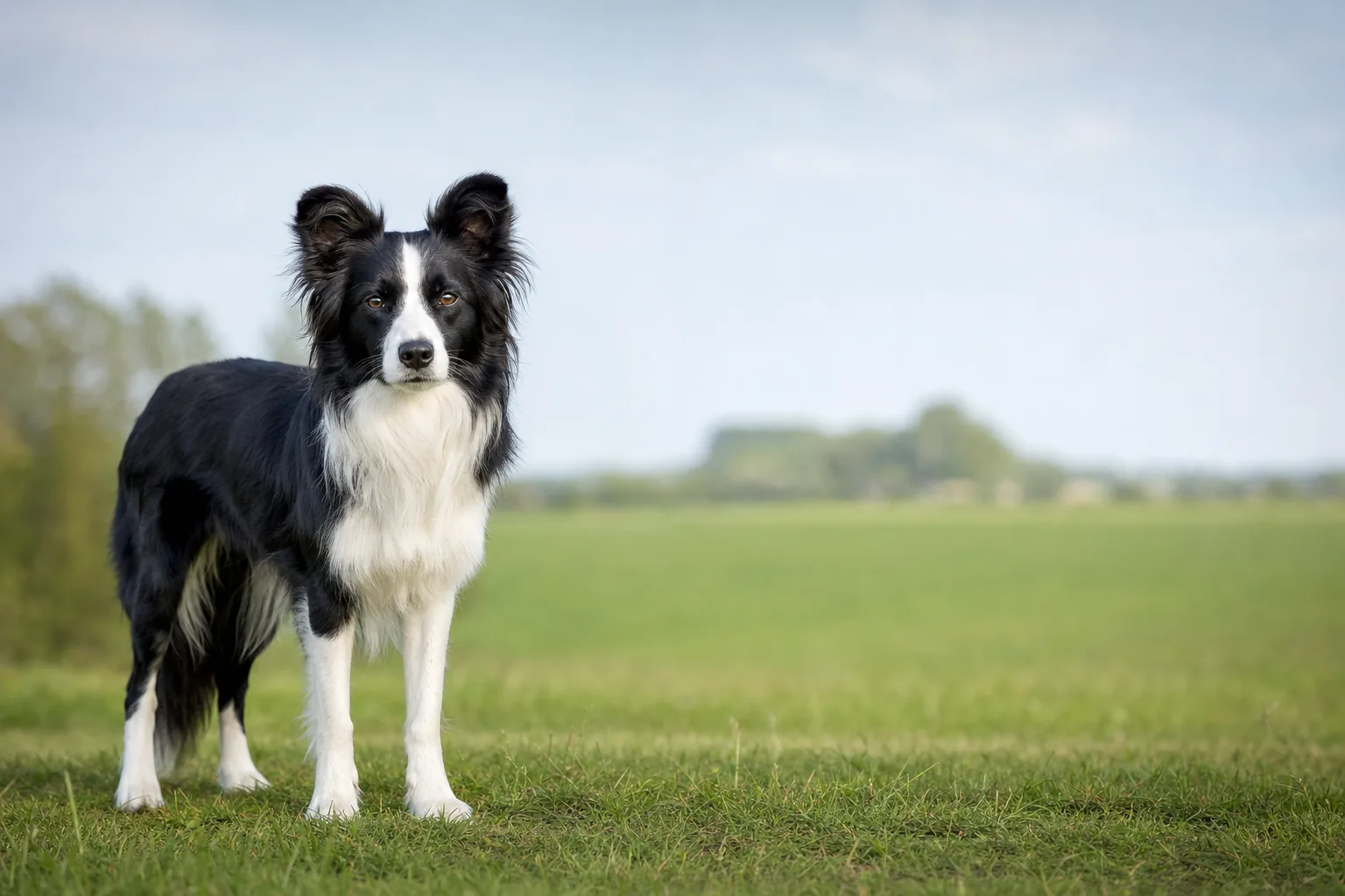
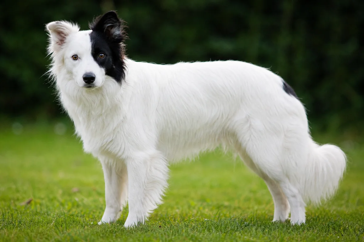
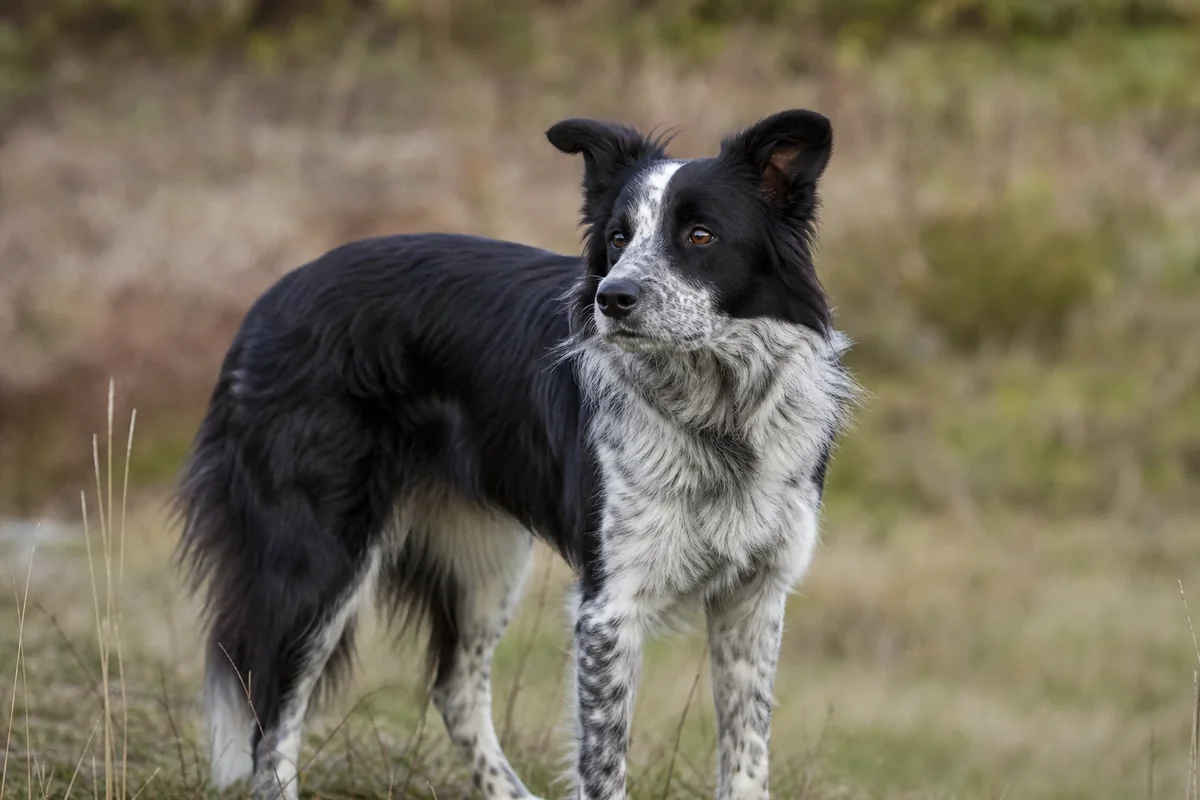
Describe the nose as black, brown, grey/slate, uniformly pink or partly pigmented, without promoting a colour name into a genotype. Stable unpigmented areas can accompany inherited markings, especially where white occurs on the face. Pigmented areas can expand or change as a puppy matures. A breeder's label, registry description or photograph still does not identify every relevant variant.
Acquired pigment loss has many possible explanations. It may occur with inflammation, trauma, immune-mediated disease, infection or other disorders; appearance alone cannot separate them. The Merck Veterinary Manual review of nasal dermatoses shows why depigmentation must be assessed alongside texture, crusts, erosions, ulcers, bleeding, swelling and changes elsewhere on the skin. Do not apply medication or supplements based only on an online colour comparison.
Stable variation versus an acquired change
A long-standing, comfortable, symmetrical appearance is different from a new or progressive change. Arrange veterinary assessment for squinting, repeated blinking, pawing, redness, discharge, cloudiness, a newly altered iris, unequal or misshapen pupils, swelling, light sensitivity, apparent difficulty seeing, or any eye that looks or feels painful. Photographs taken before the change can help establish timing, but they cannot establish the cause.
Some signs are time-critical. Sudden vision loss, a protruding eye, major trauma, a possible penetrating injury or foreign body, severe pain, rapid clouding, or an abrupt marked pupil change requires urgent veterinary guidance. The Merck Veterinary Manual overview lists acute glaucoma, lens displacement, corneal injury and acute vision loss among ophthalmic emergencies. Prevent rubbing if this can be done safely and call before travelling; do not use leftover eye drops, because treatment that suits one cause can be harmful in another.
For the nose, seek prompt care for progressive pigment loss with crusting, erosions or ulcers, persistent discharge, swelling, pain or recurrent bleeding. Breathing difficulty, severe trauma or bleeding that does not stop is urgent. These triage signs do not identify a disease—they indicate that an examination should not be replaced by a colour guide.
What an eye examination and a DNA test establish
A veterinary eye assessment starts with history and a direct examination. Depending on the concern, the clinician may assess visual responses and pupils, use magnification and ophthalmoscopy, stain the cornea, measure tear production or intraocular pressure, or recommend imaging and referral. The selection and interpretation of tests belong to the examining professional.
An OFA Companion Animal Eye Registry (CAER) screening is performed by a board-certified veterinary ophthalmologist and records observable findings at that examination. OFA explicitly notes that this screening is not a comprehensive ocular health examination: procedures such as tonometry, corneal staining, tear testing, gonioscopy or electroretinography are not routinely included. It is a dated phenotype record, not lifelong proof of health and not a DNA result. The ACVO explanation recommends repeat screening because some inherited conditions appear later.
A DNA test answers an even narrower question: whether the assayed variant was detected in the submitted sample. For example, the UC Davis Collie Eye Anomaly test targets a variant associated with one inherited disorder; it does not certify normal iris colour, exclude every cause of coloboma, or replace an eye examination. Verify the dog's permanent identity, laboratory, exact variant, method, date and report version. “Clear panel” is not a complete eye-health conclusion.
Standards and sensible records
The FCI Border Collie standard describes brown eyes, while allowing one or both eyes, or part of an eye, to be blue in merles; it also describes nose colours associated with brown and blue coats. Those are conformation descriptions, not genetic assays or clinical findings. Meeting a standard cannot prove healthy eyes, and a cosmetic departure cannot diagnose disease.
Keep observations, documents and interpretations separate. For an individual dog, retain dated photographs, veterinary findings, original laboratory reports and the dog's verified ID. Ask what was directly observed, what was tested, which variants were included, and what remains unknown. Colour cannot rank temperament, working ability, welfare or breeding quality, and this guide provides no mating rule.
Frequently asked questions
Does a blue eye prove that a Border Collie is merle?
No. Merle can be associated with blue or partly blue irises, but blue eyes have other possible genetic explanations and may occur without a visible merle coat. A photograph cannot establish PMEL genotype.
Is heterochromia always an eye disease?
No. Stable congenital complete or sectoral heterochromia can be a normal pigment variation. A new colour change, especially with pain, redness, cloudiness, pupil change or altered vision, needs veterinary assessment.
Can nose colour reveal a dog's B- or D-locus genotype?
Not by itself. Black, brown or diluted external pigment may be compatible with particular coat-colour pathways, but lighting, patterning, other variants and acquired change limit inference. Only an appropriately interpreted test documents the tested variants.
Can an eye photograph show that a dog sees normally?
No. A photograph records surface appearance under one set of conditions. It cannot assess visual pathways, intraocular pressure, the full retina or every inherited and acquired disorder.
What does a CAER eye screening establish?
It records observable ocular findings during a standardized screening by a board-certified veterinary ophthalmologist. It is time-specific, is not a DNA test and does not include every procedure used in a comprehensive diagnostic examination.
Which eye or nose signs need urgent veterinary help?
Seek urgent veterinary guidance for sudden vision loss, severe eye pain, major trauma, a protruding eye, rapid clouding, a marked pupil change, breathing difficulty or uncontrolled nasal bleeding. Redness, squinting, discharge, ulcers, crusting or progressive pigment change also warrant prompt assessment.
Sources and further reading
- Fédération Cynologique Internationale — Border Collie standard No. 297.
- UC Davis Veterinary Genetics Laboratory — dog coat-colour overview.
- UC Davis Veterinary Genetics Laboratory — brown/TYRP1 test.
- UC Davis Veterinary Genetics Laboratory — dilute/MLPH test and known limits.
- UC Davis Veterinary Genetics Laboratory — merle/PMEL test.
- Murphy et al. (2018) — PMEL insertion length and merle phenotype.
- Deane-Coe et al. (2018) — blue eyes and heterochromia association near ALX4.
- Platt et al. (2006) — congenital deafness and phenotype in Border Collies.
- Orthopedic Foundation for Animals — CAER eye screening and limitations.
- American College of Veterinary Ophthalmologists — CAER examinations.
- UC Davis Veterinary Genetics Laboratory — Collie Eye Anomaly test.
- Merck Veterinary Manual — ophthalmic emergencies in small animals.
- Merck Veterinary Manual — nasal dermatoses of dogs.
Clinical limit: this reference supports observation and preparation. It cannot diagnose an eye or nose condition, determine an individual genotype or replace veterinary care.