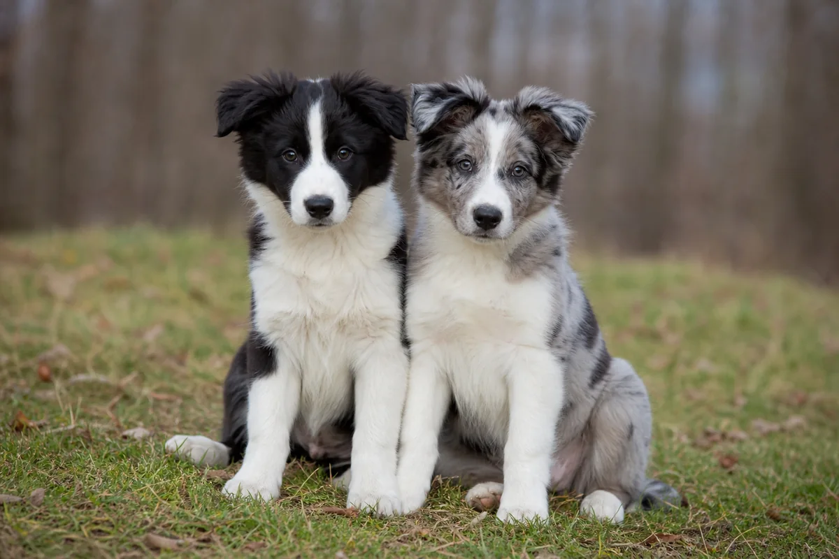
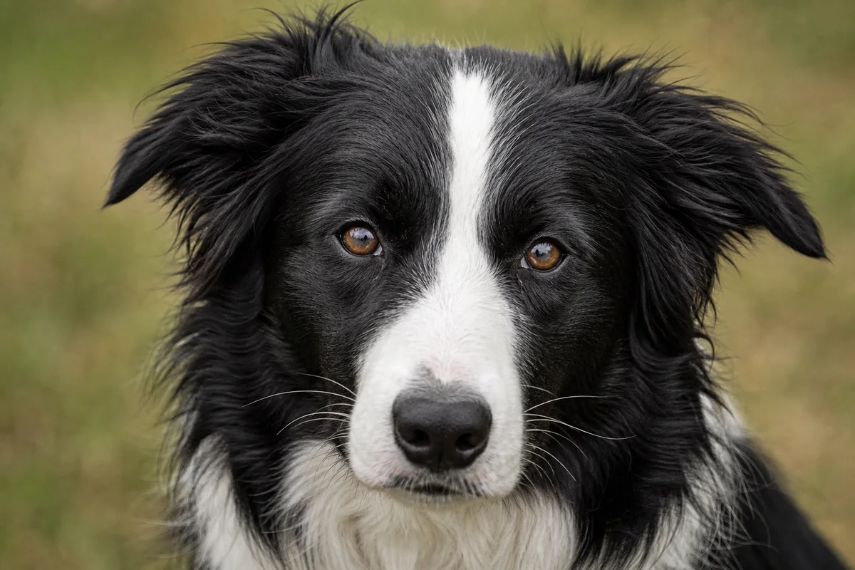
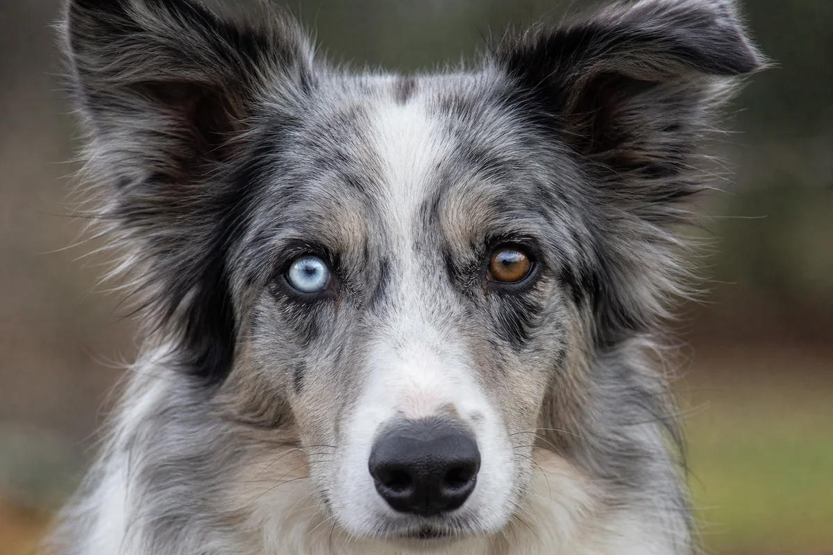
Merle is best recorded in layers. First describe the visible pattern and base pigment. Then record the laboratory's result, including insertion size and mosaic findings. Keep hearing and eye examinations separate: neither a photograph nor a DNA colour report diagnoses sensory function.
What merle looks like—and what a photograph cannot show
Merle usually creates irregular areas of lighter and full-strength eumelanin, producing a marbled or patched appearance. On a black-based coat, the familiar label is often “blue merle”; on visible brown eumelanin, “brown merle,” “chocolate merle” or “red merle” may be used. If the base is already MLPH-related dilute, communities may use “slate merle.” These names are conventions, not complete genotypes.
Other systems change what is visible. An e/e red dog may not show the expected eumelanin pattern in its hair, even if it carries a merle-associated insertion. Merling may also be limited to a tiny area or become difficult to see with age, clipping, white markings or photography. Nose and iris pigment can add descriptive clues, but neither proves an allele. The FCI Border Collie standard allows blue in one or both eyes in merles; that conformation language is not a health assessment.

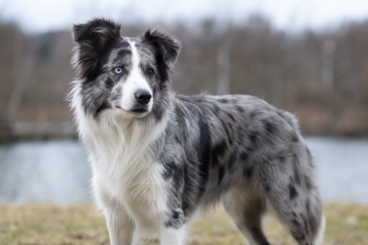
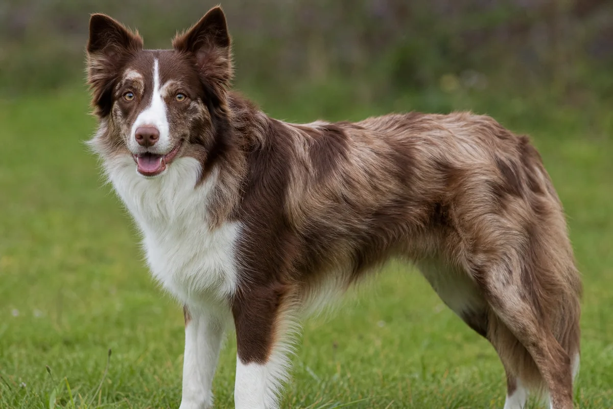
These illustrations demonstrate naming conventions only. They cannot verify base colour, PMEL allele length, mosaicism, parentage or sensory health.
PMEL and the merle-associated insertion
The locus historically called M maps to PMEL, also called SILV or PMEL17 in older literature. The landmark 2006 primary study identified a SINE retrotransposon insertion at an intron–exon boundary of the gene. PMEL contributes to the structure of eumelanin-containing pigment organelles. The insertion can alter RNA processing and pigment deposition, helping explain the characteristic patchwork.
The simple symbols m and M remain useful teaching shorthand. They are not the whole current model. A variable poly(A) tract forms part of the insertion, and its measured length correlates broadly with expression. Studies have proposed groups such as Mc, Mc+, Ma, Ma+, M and Mh, but their size boundaries and terminology are not universal. UC Davis now reports measured merle allele sizes because published naming systems differ.
Allele length is informative, not perfectly predictive
Two influential 2018 studies—one on length variation within the PMEL retrotransposon and another describing allele classes from Mc to Mh—both connect insertion length with phenotype, while using different bins and interpretations. A later review of the molecular evidence discusses those differences and the limits of detecting minor cell populations.
Accordingly, a base-pair number should be read with the laboratory's assay, reference markers, calling thresholds and current interpretation. Adjacent size ranges may overlap in appearance; other colour loci can mask expression; and the same broad label does not guarantee the same coat. A result should not be converted automatically between laboratories. Ask what the test detects, whether it sizes the insertion, how it reports multiple peaks and whether the method has been validated for the submitted sample type.
Mosaicism changes what one sample can tell you
The repetitive tract can change length during cell division. When different cell populations carry different lengths, the dog is mosaic. Somatic mosaicism can contribute to patches across the coat, while germline mosaicism can complicate inheritance. Blood, cheek cells, coat and reproductive cells do not necessarily contain identical proportions of every cell population. A low-frequency peak may also sit below an assay's detection threshold.
Why “merle never skips a generation” is too absolute
A visually plain dog may carry a short, minimally expressed or colour-masked merle-associated insertion. Mosaicism adds another layer. An unexpected pattern is a reason to verify identity, records and an appropriate laboratory report—not a basis for making allegations from appearance alone.
Keep the original report with the dog's permanent identification, sample date and laboratory name. Look for the actual allele sizes, the report's nomenclature key and any notation for mosaic peaks. A “negative” or “non-merle” headline only addresses the sequence and detection scope of that assay in that sample.
Hearing and eye risks: association is not destiny
Dogs carrying two merle-associated PMEL alleles are not one uniform category. Available evidence associates some combinations—especially two typical, strongly expressed merle alleles and extensive pigment reduction—with a higher frequency of congenital auditory and ocular abnormalities. The everyday term “double merle” can therefore obscure meaningful differences in allele length, mosaicism and phenotype. Conversely, calling a dog “single merle” is not a certificate of normal hearing or eyes.
A 2009 BAER study of 153 merle dogs found hearing loss in both heterozygous and homozygous groups, with a significant difference between them. Only five Border Collies were included, so those data cannot supply a reliable Border Collie-specific percentage. Hearing can be normal in both ears, absent in one ear or absent in both. Dogs with unilateral deafness may compensate so well that everyday observation misses it.
Reported eye abnormalities in merle-associated ocular dysgenesis include unusually small eyes, iris or other colobomas, misshapen pupils, lens defects and retinal abnormalities. Severity varies between dogs and between eyes. Evidence includes an older controlled ophthalmic study in Dachshunds and a modern veterinary case report. Those reports show possible manifestations; they do not establish that every merle dog, or every blue-eyed dog, is affected. Border Collies can also have unrelated eye disease, so visual guessing is particularly unsafe.
Screen function instead of inferring it
Hearing: the brainstem auditory evoked response, or BAER, records neurologic responses to sound for each ear. The OFA congenital-deafness protocol describes BAER as the accepted diagnostic method and requires a tracing linked to the dog. A startle response, response to clapping or apparent obedience cannot rule out unilateral deafness. Ask a veterinarian or established BAER service about appropriate timing and preparation.
Eyes: an examination by a veterinary ophthalmologist assesses observable eye structure and may indicate whether further testing is needed. The OFA eye programme stresses that screening is not a comprehensive ocular examination and reports phenotype, not hidden genotype. The American College of Veterinary Ophthalmologists explains the role of its board-certified specialists and CAER screening. Seek veterinary care promptly for squinting, redness, discharge, cloudiness, eye rubbing, a visibly small eye or pupil abnormality, or concern about navigation or vision.
Verify results and ask the right professional
For a DNA report, match the registered name and microchip or other permanent ID, sample date, laboratory, assay, allele sizes and mosaic notation. For BAER, check that both ears are identified and that the tracing, interpretation, date, examiner and dog ID belong together. For an eye result, retain the complete ophthalmologist's form rather than a cropped badge or database screenshot; note the exam date and its scope.
Questions about an individual dog's hearing, eyes or new symptoms belong with a veterinarian and, when indicated, a neurologist or veterinary ophthalmologist. Complex PMEL results may warrant a laboratory geneticist or veterinary genetics professional. Reproductive decisions require breed-relevant health, welfare, behaviour and diversity information beyond coat colour; this guide deliberately does not provide a mating formula.
Frequently asked questions
Does every merle-looking dog have the same PMEL genotype?
No. Merle-associated insertions vary in length, dogs may be mosaic, and other coat systems alter what is visible. A photograph cannot establish the complete genotype.
Can merle appear to skip a generation?
The inherited PMEL insertion does not need to produce an obvious pattern. Shorter alleles, very small merled areas, masking by another colour system and mosaicism can make a dog look non-merle. Records and an appropriate test are more reliable than appearance alone.
What do labels such as Mc, Ma, M and Mh mean?
They are proposed categories based largely on measured insertion length. Published and laboratory boundaries differ, so read the base-pair result, method and definitions on the specific report instead of translating labels automatically.
Does a blue eye prove that a merle dog is deaf or visually impaired?
No. Iris colour is not a hearing or vision test. Hearing status requires BAER testing, and eye structure and function require veterinary examination.
How is hearing assessed in a merle dog?
A BAER test records the brainstem response to sound and assesses each ear. Behavioural response alone can miss unilateral deafness, so keep the tracing and report linked to the dog's verified identity.
Can a PMEL DNA result predict an individual dog's hearing and vision?
No. It reports the merle-associated alleles detected by that assay in that sample. It does not diagnose deafness or eye disease; BAER testing and an examination by a veterinary ophthalmologist answer different clinical questions.
Sources and further reading
- Fédération Cynologique Internationale — Border Collie standard No. 297.
- UC Davis Veterinary Genetics Laboratory — Merle test and reporting.
- Clark et al. (2006) — the merle-associated SINE insertion in canine SILV/PMEL.
- Murphy et al. (2018) — length variation within the canine PMEL merle retrotransposon.
- Langevin et al. (2018) — merle phenotypes, insertion sizes and mosaicism.
- Varga et al. (2020) — review of the molecular genetics of canine merle.
- Strain et al. (2009) — BAER-assessed deafness in heterozygous and homozygous merle dogs.
- Dausch et al. (1978) — ophthalmic findings in merle Dachshunds.
- Bauer, Sandmeyer and Grahn (2015) — merle ocular dysgenesis case report.
- Orthopedic Foundation for Animals — congenital deafness and BAER protocol.
- Orthopedic Foundation for Animals — eye screening scope and interpretation.
- American College of Veterinary Ophthalmologists — CAER eye screening.
Editorial scope: this guide supports careful reading of colour and screening records. It cannot diagnose hearing or eye disease, predict an individual outcome or replace professional genetics and veterinary advice.
The discipline of reading a label rather than guessing continues in a note on how a Negroni balances bitterness, sweetness and dilution.