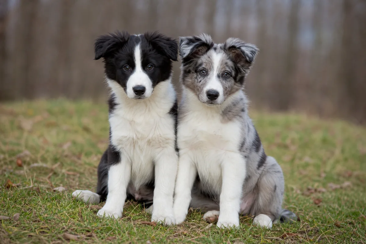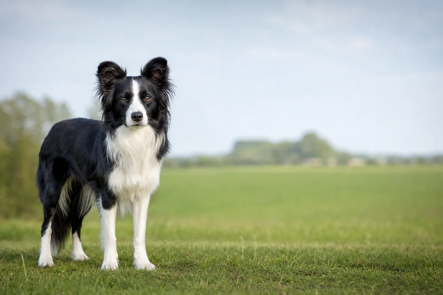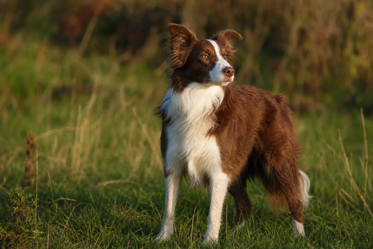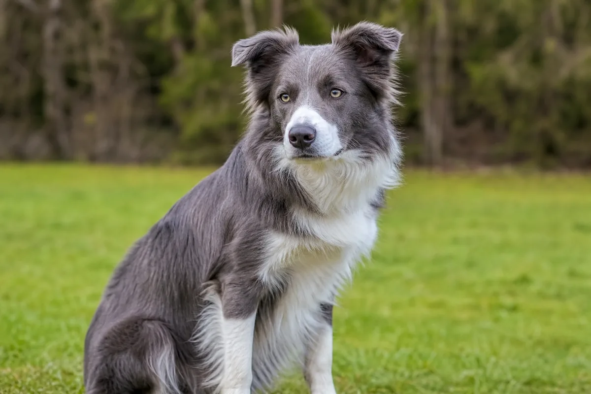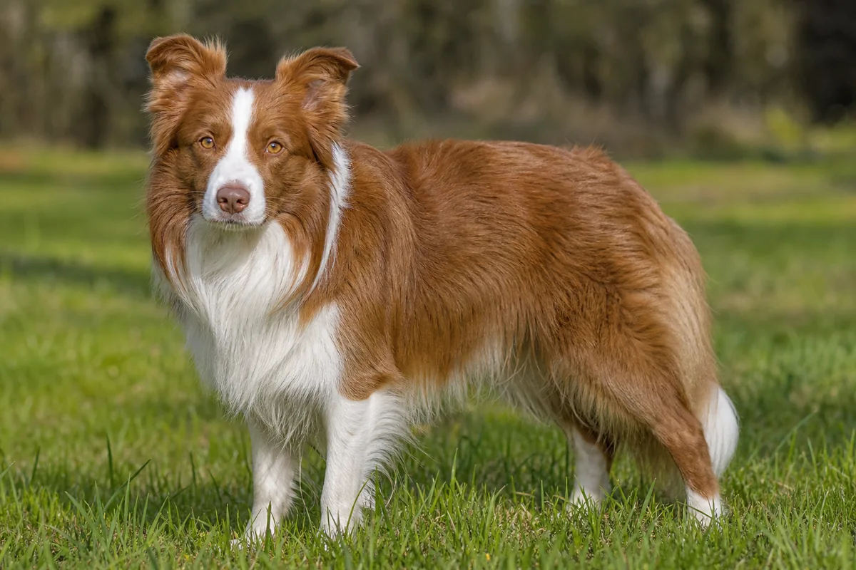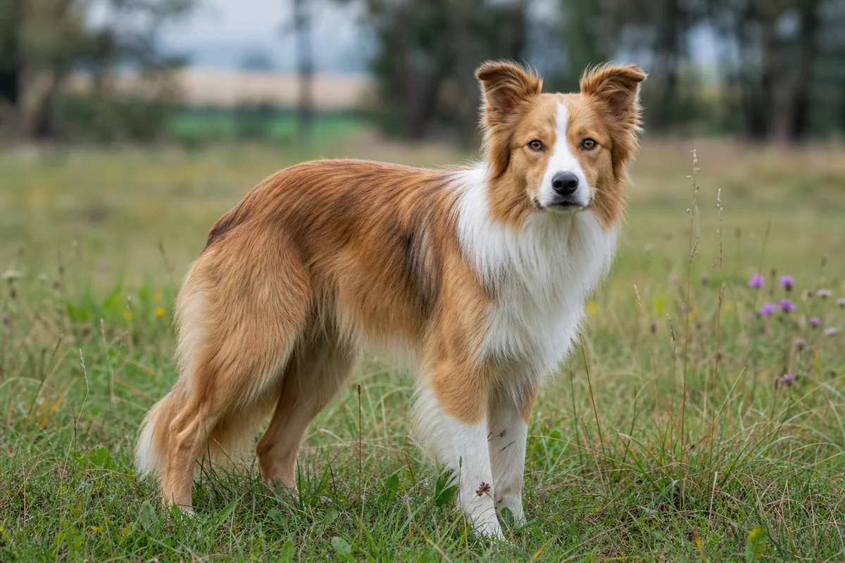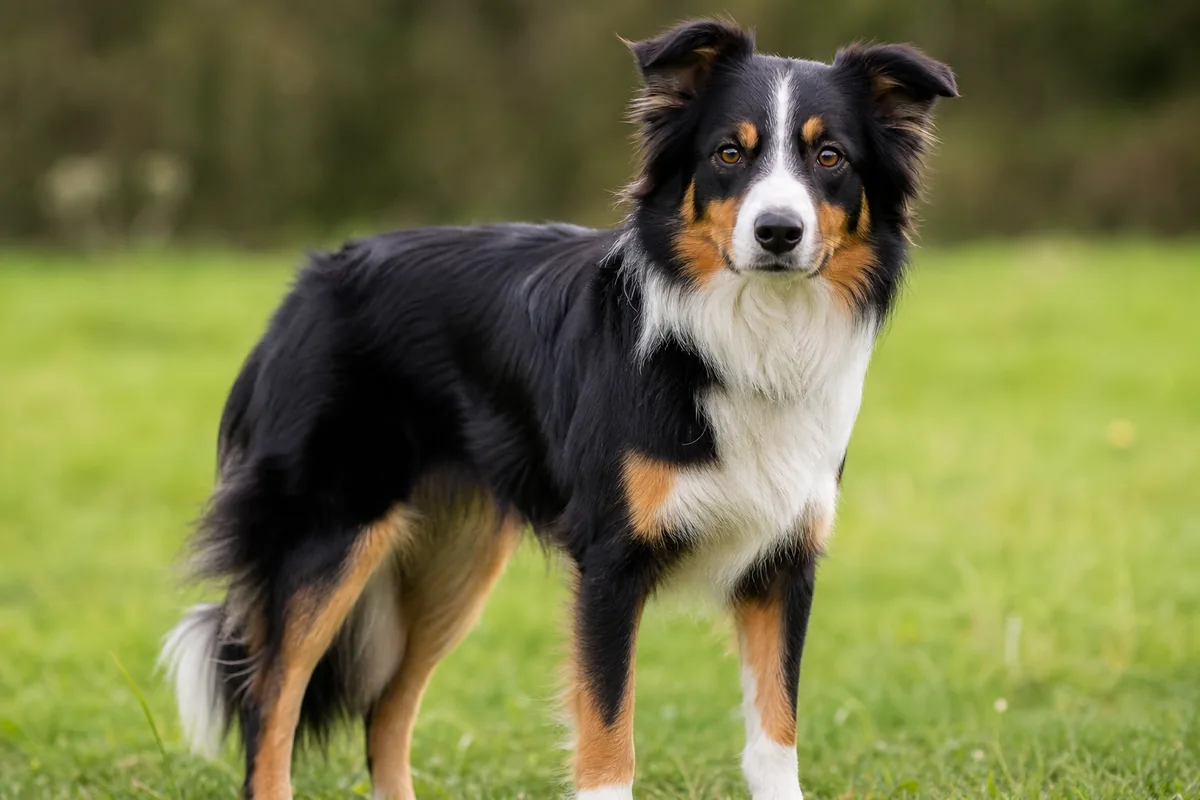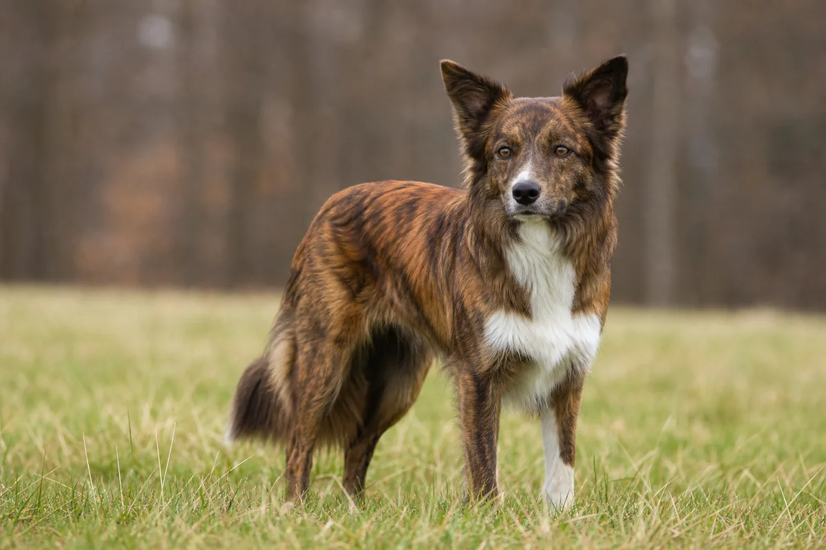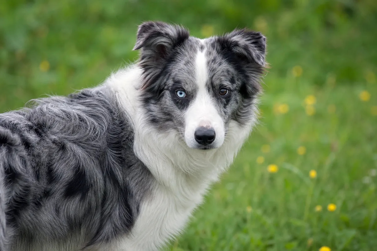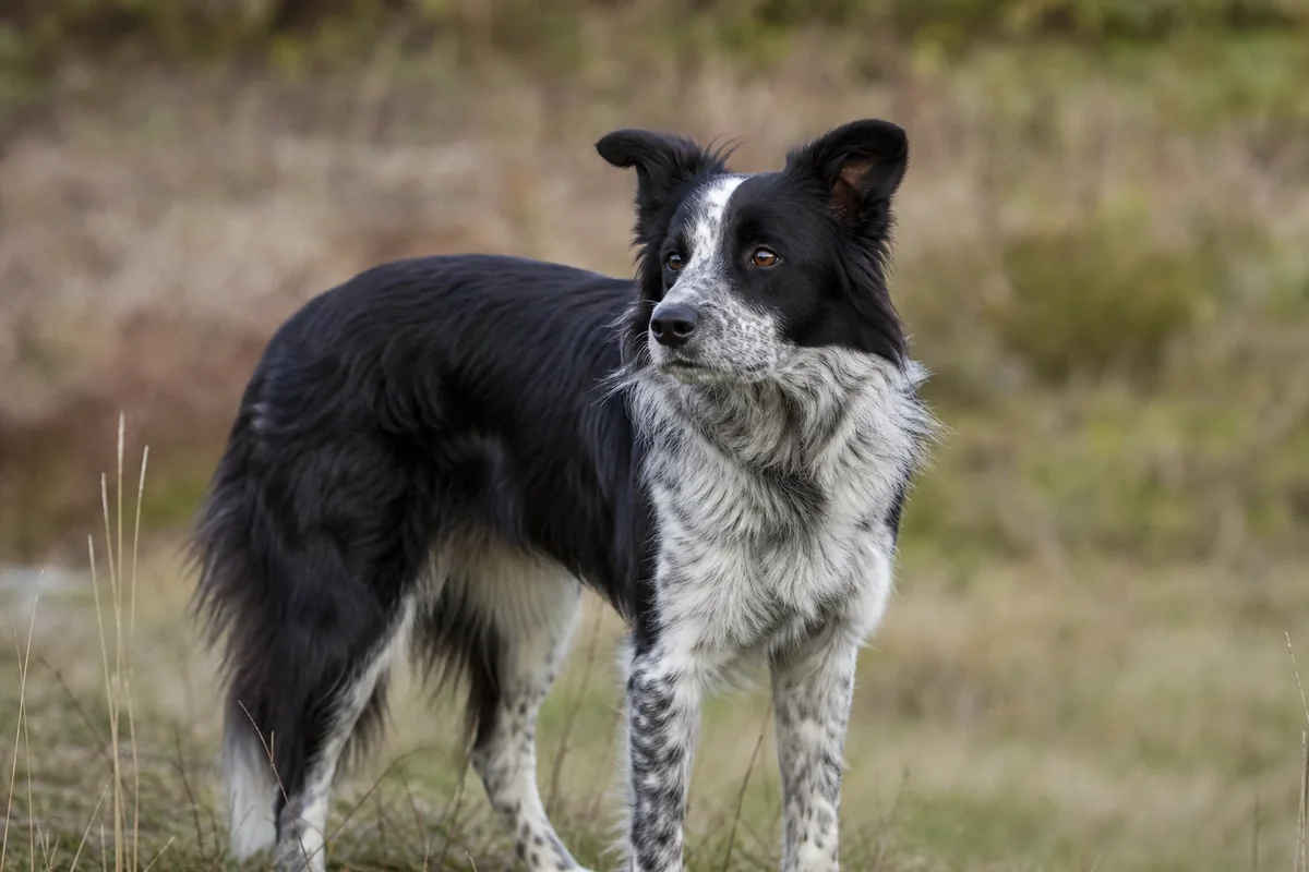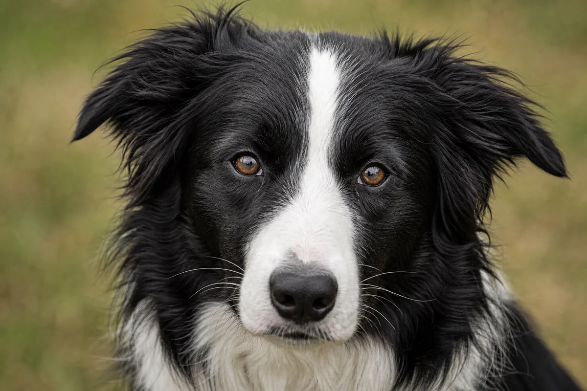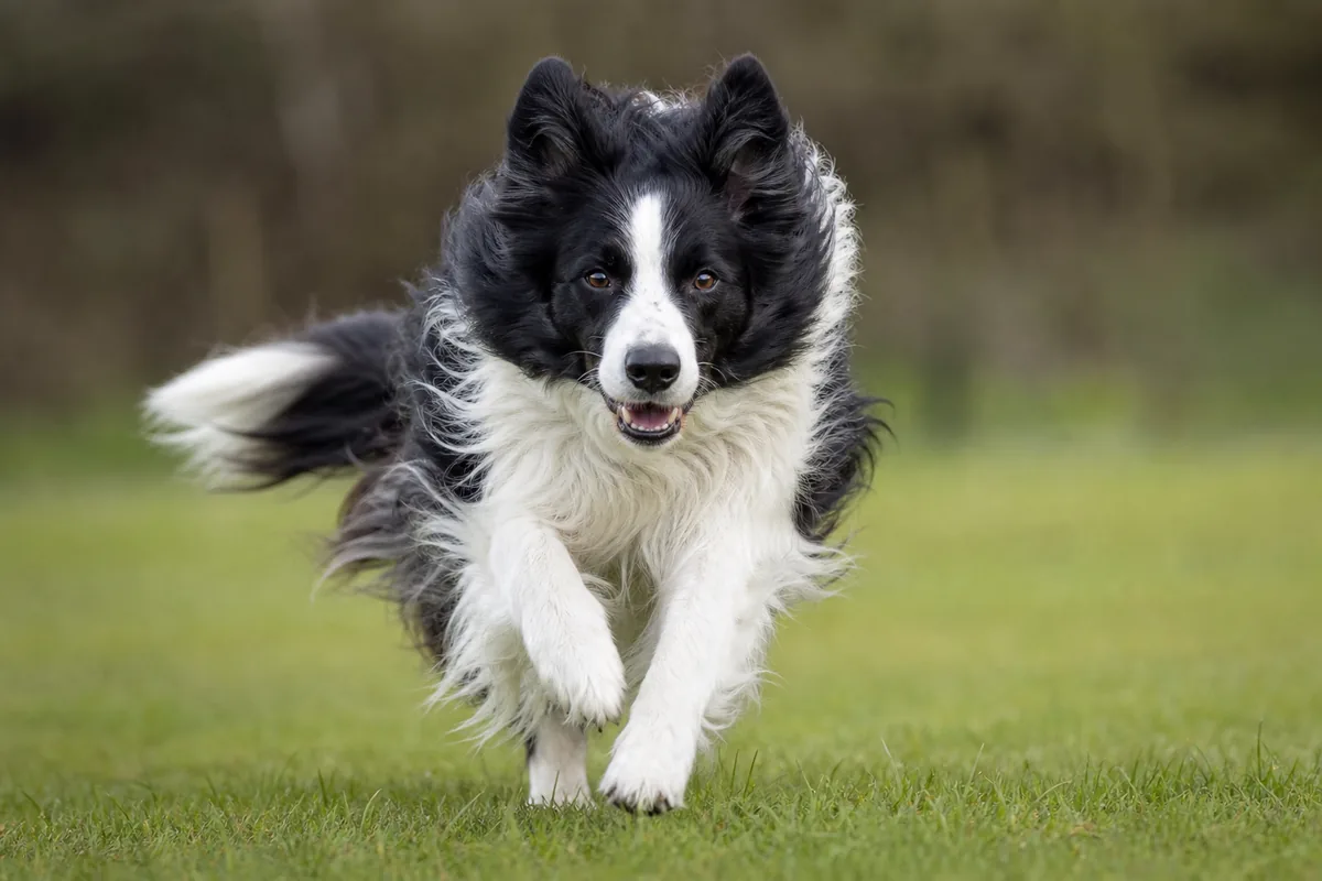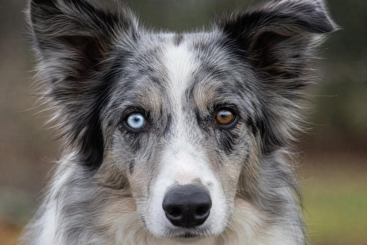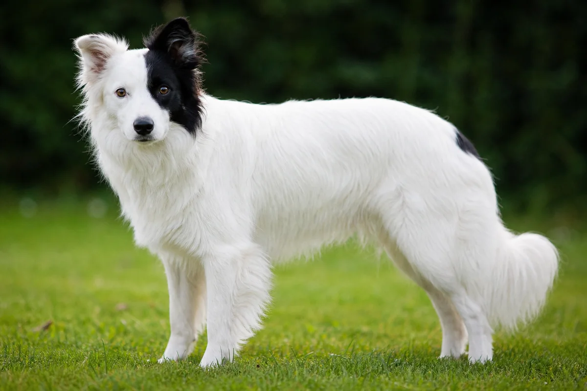
Start by describing where pigment is present and absent. Then ask which genetic mechanism a particular test covers. Keep sensory screening separate. A symmetrical collar, a mostly white body or a coloured head can guide vocabulary, but none reveals every allele or the function of an individual dog's ears and eyes.
White hair is not one biological category
In true white spotting, pigment cells are absent from affected hair follicles, so those hairs grow without pigment. “Irish spotting” usually describes a broadly symmetrical pattern involving some combination of blaze, muzzle, chest, neck, feet, underside and tail tip. “Piebald,” “parti” or “random white” often describes less symmetrical or more extensive areas. Boundaries vary, and neither label is a laboratory diagnosis.
Record the dog's age, natural-light appearance, skin and nose pigment, eye colour, and whether pale areas contain truly white hairs or very light cream. Note markings on both sides: a photograph from one angle can miss a body patch. Ticking or roaning can later add pigmented hairs inside a white area; it modifies the appearance of an existing white background rather than explaining why that area was white initially.
The historical S-locus model and modern MITF evidence
Traditional texts describe an allelic series—solid S, Irish spotting sᶦ, piebald sᵖ and extreme white sʷ. That model remains useful for discussing phenotypes, but it predates current molecular work and should not be treated as four universally testable switches.
The major canine white-spotting region contains MITF, a regulator important to pigment-cell development. A 2014 functional study of the MITF-M promoter found that repeat length affects promoter activity and concluded that familiar spotting categories are better understood through combinations of regulatory variants and haplotypes, not three independent mutations neatly matching every traditional label. Developmental variation and other modifiers also influence the final edges and extent.
Irish spotting is a phenotype first
A classic-looking collar and blaze do not prove sᶦ/sᶦ. Some “pseudo-Irish” patterns can arise from dosage of a piebald-associated haplotype, while the full genetic basis of Irish spotting is not reduced to one commercial result. Use the visual term without overclaiming the genotype.
Other routes to white or nearly white hair
Extension and pigment intensity: dogs with certain recessive MC1R variants make phaeomelanin rather than eumelanin in their coat. The resulting red, yellow or cream can be extremely pale. The UC Davis E-locus resource explains that several known MC1R variants have different breed distributions, while primary studies identify additional influences on phaeomelanin intensity. A pale e/e coat is not the same as an unpigmented MITF white patch; nose, lip and eye-rim pigment often remains.
Merle: some PMEL merle configurations can reduce eumelanin or remove visible pigment across large areas. Merle may itself be subtle or masked by e/e red, so a white-looking coat must not be assigned to MITF from appearance alone. PMEL testing answers a different question from piebald testing.
Less common or acquired change: pigment-production disorders, scarring, inflammation and immune-mediated depigmentation can also alter hair or skin. New or progressive depigmentation, hair loss, irritation or texture change belongs with a veterinarian. These are clinical questions, not names for a puppy's stable inherited markings.
What a piebald test can—and cannot—say
The UC Davis piebald/white-spotting test detects a particular MITF variant associated with piebald or random white spotting in many breeds, including the Border Collie. Its report uses N and S, where S denotes the tested piebald variant. That laboratory-specific “S” is not automatically the same symbol as solid colour in the historical four-allele model—an excellent example of why the report's legend matters.
A detected variant can support one explanation; it cannot measure every modifier, resolve all Irish spotting, identify every cause of white or predict the exact pattern. A non-detected result means that named variant was not found in that sample. It does not make the dog genetically “clear of white.” Check the dog's permanent ID, sample date, laboratory, method, exact variant, breed-validation note and panel version. Keep the full report rather than a copied headline.
Breed standards describe appearance, not health
The FCI Border Collie standard permits a variety of colours and states that white should never predominate. This is a conformation criterion. It neither identifies an MITF or PMEL genotype nor certifies hearing, vision, temperament or overall health. A dog outside a colour preference is not thereby diagnosed with disease, and a dog whose markings fit the standard is not thereby medically screened.
For accurate records, keep three columns: observed (where white hair appears), documented (the exact registry term and laboratory findings), and inferred (a hypothesis that still needs evidence). The face-markings guide helps describe distribution without turning a blaze or split face into a genetic or medical conclusion.
White pigmentation and congenital deafness
Pigment-associated congenital sensorineural deafness is documented in dogs, and melanocytes play a developmental role in the cochlea. However, “more white equals deaf” is not a diagnostic rule. Risk differs across breeds, families and mechanisms; many extensively white dogs hear normally, while a dog with ordinary markings can have hearing loss from genetic or non-genetic causes.
A large retrospective study of BAER-tested Border Collies reported statistical associations between congenital deafness and merle, blue irises and excess white on the head. Association does not prove that a particular white patch caused deafness, and the results cannot predict an individual. The broader review of deafness genetics in domestic animals also emphasizes that pigment-related mechanisms and inheritance vary. Ear colour or a response to a household sound cannot settle hearing status.
BAER and eye examinations answer different questions
BAER testing records the brainstem response to sound in each ear. It can distinguish bilateral hearing, unilateral deafness and bilateral deafness; everyday observation may miss a dog that hears with one ear. The OFA congenital-deafness protocol describes BAER as the accepted diagnostic method. Use an established veterinary or audiology provider, follow its preparation and timing instructions, and retain the tracing with the dog's verified identity, date and interpretation.
Eye and iris observations are separate. A blue iris is not proof of deafness, blindness or merle, and a brown iris is not a health certificate. The OFA eye programme explains that a specialist screening exam records observable phenotype and is not a complete genetic test or comprehensive examination. Consult a veterinarian or veterinary ophthalmologist for a visibly small or abnormal eye, unusual pupil shape, squinting, redness, discharge, cloudiness or concern about vision. The American College of Veterinary Ophthalmologists explains the role of board-certified specialists and CAER screening.
Use the evidence without turning it into a recipe
Coat records, MITF testing, PMEL testing, BAER and ophthalmic examinations answer different questions. No single result chooses a mating or predicts a litter's health. Reproductive decisions need wider breed-relevant health, behaviour, welfare and diversity information, interpreted with the relevant laboratory, veterinarian or genetics professional. This page intentionally provides no prescriptive colour pairing.
Frequently asked questions
Is all white in a Border Collie caused by the same gene?
No. MITF-related spotting is important, but pale phaeomelanin, merle-associated pigment loss and other genetic or acquired processes can also look white. The distribution and pigment context matter.
Does an Irish spotting pattern prove a specific genotype?
No. Irish spotting is a useful visual label for a broadly symmetrical pattern, not proof of one allele combination. Similar patterns can arise from different MITF haplotypes, modifiers and developmental variation.
Can a piebald DNA test explain every white marking?
No. A piebald test detects the variant named by that laboratory. It may clarify one MITF-associated mechanism, but it does not test every cause of white, every modifier or all Irish spotting.
Is a mostly white or white-headed Border Collie automatically deaf?
No. Population studies report associations between some pigmentation features and congenital deafness, but many white-marked dogs hear normally. Appearance cannot determine hearing in either ear.
What does a BAER test tell me?
BAER records the brainstem response to sound and assesses each ear separately. It can identify unilateral or bilateral deafness that everyday observation may miss, but it does not explain every possible cause.
Do blue eyes or breed-standard markings prove eye health?
No. Iris colour and breed-standard descriptions are not eye examinations. A veterinarian or veterinary ophthalmologist must assess structure and function when screening is needed or a concern is present.
Sources and further reading
- Fédération Cynologique Internationale — Border Collie standard No. 297.
- UC Davis Veterinary Genetics Laboratory — piebald/white spotting test.
- Baranowska Körberg et al. (2014) — MITF-M promoter repeat and white spotting.
- UC Davis Veterinary Genetics Laboratory — MC1R red/cream variants.
- Hédan et al. (2019) — phaeomelanin dilution and white or cream coats.
- UC Davis Veterinary Genetics Laboratory — merle test and allele-size reporting.
- Murphy et al. (2018) — PMEL merle insertion length and phenotype.
- Platt et al. (2006) — congenital deafness and phenotype in Border Collies.
- Strain (2015) — genetics of deafness in domestic animals.
- Orthopedic Foundation for Animals — congenital deafness and BAER protocol.
- Orthopedic Foundation for Animals — eye screening scope and interpretation.
- American College of Veterinary Ophthalmologists — CAER eye screening.
Editorial scope: this reference supports careful description and record-checking. It cannot diagnose a dog, predict an individual sensory outcome or replace veterinary or genetics advice.